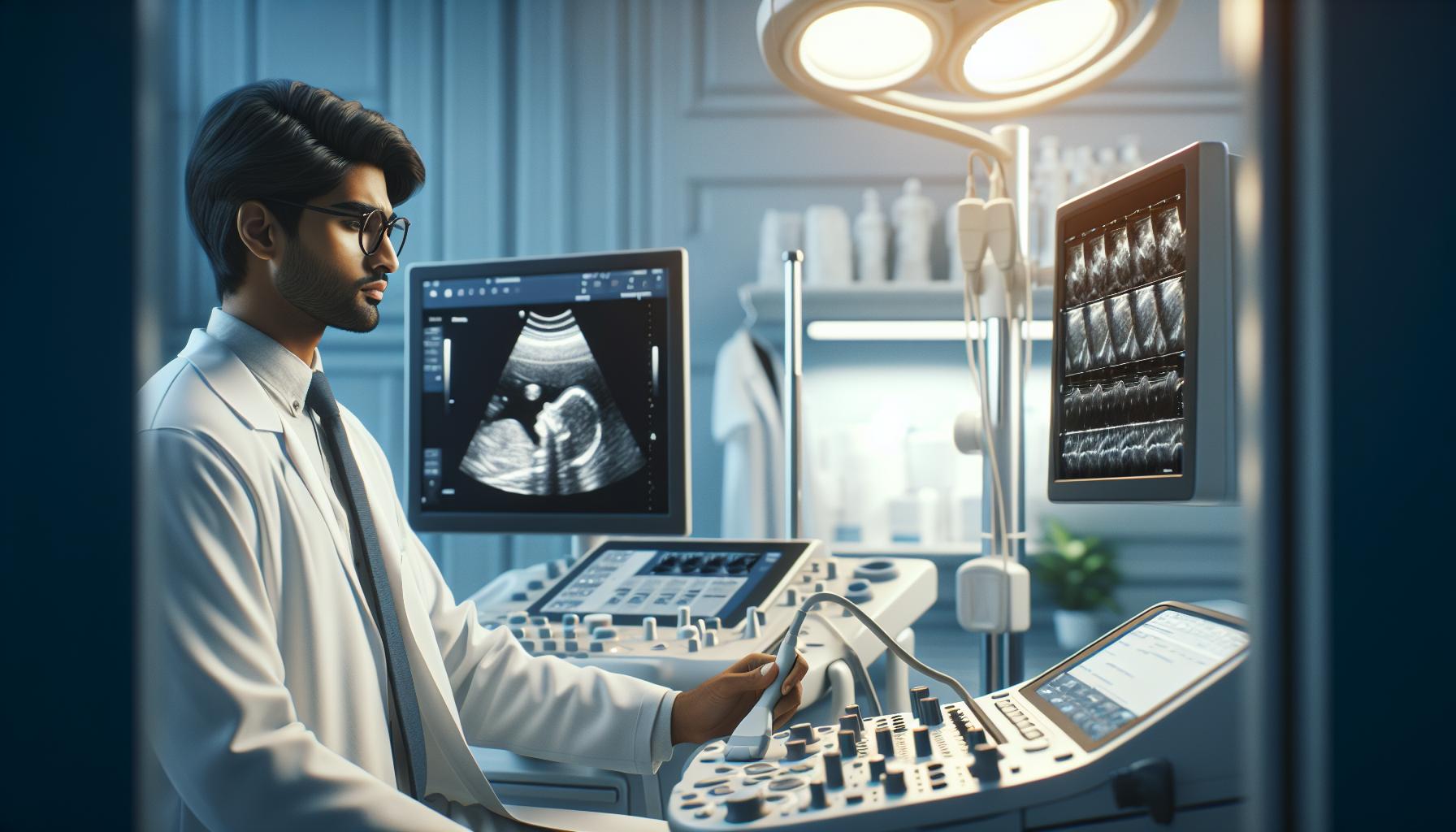
Many people wonder whether ultrasound technology and sonography refer to the same medical imaging technique. While these terms are often used interchangeably in healthcare settings, understanding their subtle differences and similarities can help clarify their roles in modern medicine. Both ultrasound technology and sonography involve using high-frequency sound waves to create detailed images of internal body structures. These non-invasive diagnostic tools have revolutionized medical imaging, allowing healthcare providers to examine organs, track fetal development, and diagnose various conditions without radiation exposure. The main distinction often lies in how these terms are used professionally rather than in their technical applications.
Is Ultrasound Tech and Sonography the Same Thing
Ultrasound technology creates medical images using high-frequency sound waves that bounce off internal body structures. These sound waves operate at frequencies between 2-18 MHz, generating real-time images called sonograms.
| Component | Function | Application |
|---|---|---|
| Transducer | Emits sound waves | Image generation |
| Processor | Converts echoes to images | Data interpretation |
| Display | Shows visual output | Diagnostic viewing |
The imaging process involves three key elements:
- Transmitting sound waves through a transducer probe
- Recording the echoes as they bounce off tissues
- Converting these echoes into digital images
Sonography represents the procedural application of ultrasound technology, encompassing:
- Diagnostic imaging of organs
- Monitoring fetal development
- Evaluating blood flow
- Guiding medical procedures
Medical professionals use specific ultrasound types for different examinations:
- Doppler ultrasound for blood flow assessment
- 3D imaging for structural analysis
- 4D ultrasound for real-time motion studies
- Elastography for tissue stiffness measurement
The technical equipment includes specialized features:
- Variable frequency transducers
- Digital image optimization
- Advanced signal processing
- Real-time image reconstruction
- Cardiovascular structures
- Musculoskeletal tissues
- Abdominal organs
- Reproductive systems
The Definition and History of Both Terms

Ultrasound technology and sonography emerged from pioneering research in sound wave applications during the early 20th century. These terms developed distinct historical paths while maintaining interconnected roles in medical diagnostics.
Origins of Ultrasound Technology
The foundation of ultrasound technology traces back to 1794 when Lazzaro Spallanzani discovered echolocation in bats. Key developments include:
- Paul Langevin created the first ultrasonic submarine detector in 1915
- Dr. Karl Dussik performed the first medical ultrasound in 1942
- Dr. George Ludwig developed the A-mode ultrasound for detecting gallstones in 1949
- Dr. John Wild introduced the B-mode scanner for tissue imaging in 1951
| Year | Innovation | Impact |
|---|---|---|
| 1915 | SONAR development | First practical ultrasound application |
| 1942 | Medical ultrasound | Brain imaging breakthrough |
| 1949 | A-mode scanning | Gallstone detection advancement |
| 1951 | B-mode imaging | Real-time tissue visualization |
- Introduction of real-time imaging systems in 1965
- Establishment of the American Society of Ultrasound Technical Specialists in 1970
- Development of color Doppler imaging in 1979
- Integration of 3D imaging capabilities in 1986
- Implementation of digital processing techniques in 1990
| Decade | Advancement | Clinical Application |
|---|---|---|
| 1960s | Real-time scanning | Dynamic organ imaging |
| 1970s | Professional recognition | Standardized practices |
| 1980s | 3D capabilities | Enhanced diagnostic accuracy |
| 1990s | Digital integration | Improved image quality |
Technical Aspects and Equipment
Modern ultrasound technology incorporates advanced digital systems and specialized equipment to produce high-quality diagnostic images. The technical components work together to create detailed visualizations of internal body structures through sound wave manipulation.
Ultrasound Machines and Tools
Ultrasound systems consist of five primary components:
- Transducer probes (2-18 MHz frequency range) for different scanning applications
- Digital beam-former for signal processing and focus control
- Central processing unit for image reconstruction
- Display monitor with high-resolution capabilities
- Data storage systems for image archiving
Essential tools include:
- Linear array transducers for superficial imaging
- Curved array transducers for deep tissue scanning
- Phased array transducers for cardiac examinations
- Color Doppler modules for blood flow assessment
| Component Type | Frequency Range | Primary Use |
|---|---|---|
| Linear Array | 7-18 MHz | Vascular, breast, musculoskeletal |
| Curved Array | 2-5 MHz | Abdominal, obstetric |
| Phased Array | 2-4 MHz | Cardiac, transcranial |
Imaging Processes
The ultrasound imaging process follows four distinct steps:
- Pulse generation through piezoelectric crystals
- Sound wave transmission into body tissues
- Echo reception from reflecting structures
- Digital conversion of echoes into visual displays
- Harmonic imaging for enhanced contrast resolution
- Speckle reduction for improved image clarity
- Compound imaging for reduced artifacts
- Tissue Doppler imaging for motion analysis
| Process Feature | Enhancement Type | Clinical Benefit |
|---|---|---|
| Harmonic Imaging | Contrast | Better tissue differentiation |
| Compound Imaging | Resolution | Reduced noise artifacts |
| Doppler Processing | Motion Detection | Accurate flow assessment |
Professional Roles and Responsibilities
Professional responsibilities in medical imaging vary between ultrasound technicians and sonographers, with distinct roles emerging from their specialized training and certifications. Each position requires specific skill sets and knowledge bases that contribute to diagnostic imaging excellence.
Ultrasound Technician’s Scope of Work
Ultrasound technicians focus on operating and maintaining imaging equipment. Their core responsibilities include:
- Operating ultrasound machines with precision for basic imaging procedures
- Performing routine maintenance checks on imaging equipment
- Maintaining equipment records and scheduling repairs
- Following standardized imaging protocols
- Recording basic patient data and technical parameters
- Assisting healthcare providers with image acquisition
- Processing and storing ultrasound images
- Implementing safety protocols for equipment operation
- Conducting detailed patient assessments before procedures
- Selecting appropriate imaging protocols based on patient conditions
- Performing complex diagnostic examinations across multiple specialties
- Analyzing image quality and ensuring diagnostic accuracy
- Creating detailed technical reports for physicians
- Identifying abnormal pathological conditions in images
- Collaborating with radiologists and specialists
- Training junior staff and students
- Participating in quality improvement initiatives
- Maintaining certification through continuing education
| Role Comparison | Ultrasound Technician | Sonographer |
|---|---|---|
| Education Level | Associate Degree | Bachelor’s Degree |
| Certification | Basic ARDMS | Advanced ARDMS Specialties |
| Typical Salary Range | $45,000-$65,000 | $65,000-$95,000 |
| Decision-Making Authority | Limited | Extensive |
| Specialization Options | General | Multiple Subspecialties |
Educational Requirements and Certification
Becoming a qualified ultrasound technologist or sonographer requires specific educational credentials and professional certifications. The path includes formal education through accredited programs and obtaining necessary licenses to practice.
Training Programs and Degrees
Aspiring ultrasound professionals complete specialized education through accredited institutions:
- Associate Degree programs span 18-24 months covering anatomy physiology diagnostic principles
- Bachelor’s Degree programs extend 4 years incorporating advanced imaging techniques research methods
- Certificate programs last 12-18 months designed for healthcare professionals transitioning to sonography
- Clinical training requires 1,000+ supervised scanning hours in hospital or outpatient settings
- Specialized tracks focus on specific areas like cardiac vascular obstetric or musculoskeletal imaging
| Program Type | Duration | Clinical Hours Required |
|---|---|---|
| Certificate | 12-18 months | 1,000 hours |
| Associate Degree | 18-24 months | 1,500 hours |
| Bachelor’s Degree | 4 years | 2,000 hours |
- ARDMS certification through passing Sonography Principles & Instrumentation examination
- Specialty examinations in areas like abdomen breast cardiac obstetrics
- State-specific licensing requirements vary by region jurisdiction
- Continuing education credits maintain active certification status
- Registry credentials include RDMS RVT RDCS based on specialization
| Certification Type | Requirements | Renewal Period |
|---|---|---|
| ARDMS | SPI + Specialty Exam | Every 2 years |
| ARRT | Primary + Post-Primary | Annual |
| CCI | Qualifying Exam | Every 3 years |
Career Opportunities and Specializations
Diagnostic medical sonography offers diverse career paths across multiple healthcare settings:
Clinical Specializations:
- Obstetric and Gynecological Sonography: Examines female reproductive systems and monitors fetal development
- Abdominal Sonography: Images organs like liver, kidneys, pancreas, spleen
- Cardiac Sonography: Evaluates heart structures and blood flow patterns
- Vascular Sonography: Assesses blood vessels and circulation
- Neurosonology: Images brain and nervous system structures
- Musculoskeletal Sonography: Examines muscles, tendons, joints, ligaments
Work Environments:
- Hospitals (48% employment rate)
- Private diagnostic centers (22% employment rate)
- Physicians’ offices (19% employment rate)
- Outpatient care facilities (7% employment rate)
- Mobile imaging services (4% employment rate)
| Career Advancement Path | Average Salary Range (USD) | Years of Experience |
|---|---|---|
| Entry-Level Sonographer | $55,000 – $65,000 | 0-2 years |
| Senior Sonographer | $70,000 – $85,000 | 3-5 years |
| Lead Sonographer | $85,000 – $95,000 | 5-10 years |
| Department Supervisor | $90,000 – $110,000 | 10+ years |
- Clinical applications specialist
- Ultrasound department manager
- Education program director
- Research sonographer
- Equipment sales representative
- Quality assurance coordinator
Each specialization requires specific ARDMS certifications with documented clinical hours in the chosen field. Additional credentials enhance career advancement opportunities through organizations like SDMS CCI.
While ultrasound technology and sonography are deeply intertwined they represent different aspects of the same medical imaging field. The technology provides the foundation through sophisticated equipment and sound wave manipulation while sonography encompasses the skilled application and interpretation of these tools. Understanding this relationship helps aspiring healthcare professionals choose their career paths more effectively. Whether pursuing a role as an ultrasound technician or sonographer both paths offer rewarding opportunities in the growing field of medical diagnostics. The continuous advancement of technology coupled with increasing demand for diagnostic imaging services ensures a bright future for professionals in both specialties.


